Simulation
Diffusion Simulation
Project Description
Our diffusion simulation tool (DIFSIM) is an environment for modeling and testing an entire diffusion MRI experiment. We model diffusion of water within complex geometric structures with properties similar to neural tissue, keeping track of the effects of the magnetic field gradients applied in a simulated diffusion MRI experiment, and report the resulting signal.
DIFSIM provides an environment for testing hypotheses about disease models (how changes to tissue structure or properties affect the signal), testing new pulse sequences (how different imaging techniques perform on model tissues), and developing new post-processing and analysis techniques (how the complex output can be represented in simple but meaningful ways).
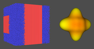
With this powerful and flexible technology, we are able to model arbitrarily complex tissues represented by triangulated meshes and can vary characteristics such as concentrations and diffusion coefficients for different regions (allowing, e.g., distinct concentrations and diffusion coefficients for intraaxonal regions, myelin sheathing, and extraaxonal fiber path material).
Publications
Berry DB, Rodriguez-Soto AE, Englund EK, Shahidi B, Parra C, Frank LR, Kelly KR, Ward SR. Multiparametric MRI characterization of level dependent differences in lumbar muscle size, quality, and microstructure. JOR Spine 2020, e1079. [Epub ahead of print].
Berry DB, Regner B, Galinsky V, Ward SR, Frank LR. Relationships between tissue microstructure and the diffusion tensor in simulated skeletal muscle. Magn Reson Med 80(1): 317-328, 2018.
Baxter GT, Frank LR. A computational model for diffusion weighted imaging of myelinated white matter. Neuroimage, 75C: 212-220, 2013.
Balls GT, Frank LR. A simulation environment for diffusion weighted MR experiments in complex media. Magn Reson Med, 62: 771-8, 2009.