The Center for Scientific Computation in Imaging (CSCI) at UCSD is
devoted to the development of addressing this critical junction
between advanced scientific instrumentation and practical quantitative
application through the development of novel principled theoretical
approaches to the spatial and temporal exploration of scientific
imaging data, and the development of efficient and accurate computational
implementations of these new approaches. Our research mission is to
provide cutting edge integrated analysis and visualization tools that
are as easy to use as they are computationally sophisticated in order
to facilitate the quantitative analysis of imaging data across a wide
range of basic scientific and clinical disciplines. This basic research
is supplemented by an educational mission to expand the understanding
of data acquisition, analysis, and visualization to the formal
education of students and researchers across a wide and ever expanding
scope of scientific investigation, while providing the computational
tools to both learn and apply these methods in their work. In addition,
our public outreach is aimed at educating the public in the importance
and utility of basic research in a wide range of application that have
a direct impact on their daily lives, and motivating an interest in
science among younger students, particularly in underserved populations,
by demonstrating the fascinating world of imaging in science.
Publications
Galinsky VL, Frank LR. Symplectomorphic registration with phase space
regularization by entropy spectrum pathways. Magn Reson Med
(accepted), 2018.
Galinsky VL, Paulus M, Martinez A, Frank LR. Joint estimation of
effective brain wave activation modes using EEG/MEG sensor arrays and
multimodal MRI volumes. Neural Comput30(7): 1725-1749, 2018.
Galinsky VL, Frank LR. A unified theory of neuro-MRI data shows scale–free nature of connectivity modes. Neural Comput 29(6): 1441-1467, 2017. doi: 10.1162/NECO_a_00955
Frank LR, Galinsky VL. Detecting spatio-temporal modes in multivariate data by entropy field decomposition. J Phys A49: 395001, 2016.
Frank LR, Galinsky VL. Dynamic multiscale modes of resting state brain activity detected by entropy field decomposition. Neural Comput, 28(9): 1769-1811, 2016.
Galinsky VL, Frank LR. The lamellar structure of the brain fiber pathways. Neural Comput, 28: 2533-2556, 2016.
Galinsky VL, Frank LR. Simultaneous multi-scale diffusion estimation and tractography guided by entropy spectrum pathways. IEEE Trans Med Imaging, 34: 1177-1193, 2015.
Frank LR, Galinsky VL. Information pathways in a disordered lattice. Phys. Rev. E.89: 032142, 2014.
Galinsky VL, Frank LR. Automated segmentation and shape characterization of volumetric data. Neuroimage92: 156-168, 2014.
CSCI has a long history of collaborations in clinical neuroscience
with neuroimaging research focused on the neurological impacts of
substance abuse and neurodegenerative diseases, such as traumatic
brain injury (TBI) and mild cognitive impairment (MCI), and
Alzheimer's disease (AD). Our most recent NIH-funded collaboration
(R01AG054049), with Co-PI Dr. Mark Bondi in Psychiatry, is utilizing
our QUEST-NI analysis and visualization methods to evaluate newly
developed diagnostic and MRI metrics for measuring changes in an area
of the brain where the first visible pathologic changes resulting from
the onset of AD are believed to occur. This new approach has the
potential to improve diagnostic and imaging precision in Preclinical
AD and impact the prospective design of future studies, as well as
promote new biomarker assessment modalities and fundamental shifts in
therapeutic targets for AD.
Publications
Sorg SF, Schiehser DM, Bondi MW, Luc N, Clark AL, Jacobson MW, Frank LR, Delano-Wood L. White matter microstructural compromise is associated with cognition but not posttraumatic stress disorder symptoms in military veterans with traumatic brain injury. Journal of Head Trauma Rehabilitation 31: 297-308, 2016.
Sorg SF, Delano-Wood L, Luc N, Schiehser DM, Hanson KL, Nation, DA, Lanni E, Jak AJ, Lu K, Meloy MJ, Frank LR, Lohr JB, Bondi MW. White matter integrity in veterans with mild traumatic brain injury: Associations with executive function and loss of consciousness. Journal of Head Trauma Rehabilitation 29: 21-32, 2014.
Delano-Wood L, Stricker NH, Sorg SF, Nation DA, Jak AJ, Woods SP, Libon DJ, Delis DC, Frank LR, Bondi MW. Posterior cingulum white matter disruption and its associations with verbal memory and stroke risk in mild cognitive impairment. Journal of Alzheimers Disease 29: 589–603, 2012.
Krysl P, Bondi MW, Ward S, Frank LR. On sources of error in finite element simulation of blast effects in the human brain. Journal of Computational Nonlinear Dynamics 7: 031008, 2012.
Working in conjunction with Dr Benjamin Zahneisen at Stanford
University, we have developed a highly efficient multi band pulse
sequence capable of acquiring multiple types of diffusion weighted
(DW) MRI data, including high angular resolution single pulsed field
gradient (sPFG) data, double pulsed field gradient (dPFG), and
hyperecho sPFG. The pulse sequence is based on the highly efficient
Extended Hybrid-Space SENSE for EPI which
provides highly efficient off-resonance and eddy current correction
using a joint interleaved blip-up/down reconstruction. This
acquisition is significantly faster than the standard acquisition that
require two (blip up & blip down) acquisitions, and also significantly
enhances image quality as the distortions are corrected as part of the
reconstruction process, rather than ex post facto. The resulting
acquisition is not only significantly more time efficient, but shows a
dramatic improvement in DWI image quality. This pulse sequence is
currently being used in a range of basic and clinical research
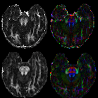
projects. Figure 1, on the right, shows FA maps (left) and directionally colored FA maps (right) from old (top) and new (bottom) pulse sequences. Marked improvement is evident.
3D DTI
High resolution, low distortion DTI remains one of the major
challenges in MR pulse sequence development. In order to reduce the
deleterious effects of the significant physiological motion in the
human brain, single shot, slice selective (i.e., 2D) EPI has been
widely adopted as the standard imaging sequence for human in vivo
diffusion MRI. However, this limits the attainable spatial resolution,
and is prone to susceptibility artifacts that significantly distort
the images. In an earlier NIH-funded project (R01 MH064729), we
developed methods for high spatial and angular resolution diffusion
tensor imaging (DTI) in humans that overcomes these limitations in
existing methods. We developed a novel high resolution, low distortion
3D Variable Density Spiral (VDS) Fast Spin Echo (FSE) diffusion tensor
imaging (DTI) pulse sequence that allows higher spatial and angular
resolution DTI and is able to image in regions of the human brain
previously unattainable due to susceptibility distortions.
Publications
Frank LR, Jung Y, Inati S, Tyszka JM, Wong EC. High efficiency,
low distortion, 3D diffusion tensor imaging with variable density
spiral fast spin echoes (3D DW VDS RARE). Neuroimage, 49:
1510-23, 2010.
Tyszka JM, Frank LR. High-field diffusion MR histology:
Image-based correction of eddy current ghosts in diffusion-weighted
rapid acquisition with relaxation enhancement (DW-RARE).
Magn Reson Med, 61: 728-33, 2009.